Wednesday, October 5, 2011

Code Blue..CPR

http://upload.wikimedia.org/wikipedia/commons/thumb/5/58/CPR_training-04.jpg/230px-CPR_training-04.jpg 
code blue team
Saving Lives at a Moment's Notice


"Code Blue, ER. Code Blue, ER."
When these words echo through Deaconess Hospital, a team of specialists drops everything and races to the Emergency Department – or wherever they are needed.


Code Blue is announced when a patient is unresponsive, meaning he is not breathing or his heart has stopped beating. When this happens, no matter what time of day or what day of the week, our code blue team is ready.


Meet Our Code Blue Team
Our team consists of the following medical professionals:


Emergency Department Physician - A board certified emergency physician oversees the code blue process.
Team Coordinator - A registered nurse from the Cardiovascular Medical Intensive Care Unit (CVICU) acts as Team Coordinator. This person is certified in advanced cardiac life support (ACLS), and is responsible for the patient’s care during a code blue.
Recorder - The recorder is an ACLS certified registered nurse from the Cardiovascular Care Center (CVCC). This team member monitors the patient throughout the process and documents the time and details of each action taken.


Medication Nurse - The medication nurse comes from the Neuro Intensive Care Unit and is also ACLS certified. This individual establishes an IV and administers medications necessary to restore a patient's vital functions.
Other Professionals - In addition to the core team described above, a pharmacist, laboratory technologist, respiratory therapist and other physicians typically assist.

During the normal course of a day, members of our code blue team are dispersed throughout the hospital, performing a variety of jobs. But when called upon, they come together and work as one unit to save yet another life.
======================

JOIN OUR PAGE IN FACEBOOK

ACLS Megacode Guildlines Treating Ventricular Fibrillation

Ventricular fibrillation (VF) begins as a quasiperiodic reentrant pattern of excitation in the ventricles with resulting poorly synchronized and inadequate myocardial contractions. The heart consequently immediately loses its ability to function as a pump. As the initial reentrant pattern of excitation breaks up into multiple smaller wavelets, the level of disorganization increases. Sudden loss of cardiac output with subsequent tissue hypoperfusion creates global tissue ischemia; brain and myocardium are most susceptible. VF is the primary cause of sudden cardiac death (SCD).

Sudden cardiac death can be viewed as a continuum of electromechanical states of the heart: ventricular tachycardia (VT), ventricular fibrillation (VF), pulseless electrical activity (PEA), and asystole. VF is the most common initial state, and, because of insufficient perfusion of vital cardiac tissues, it degenerates to asystole if left untreated.  
Treatment:


  • Defibrillation

    • Electrical external defibrillation remains the most successful treatment of ventricular fibrillation (VF). A shock is delivered to the heart to uniformly and simultaneously depolarize a critical mass of the excitable myocardium. The objective is to interfere with all reentrant arrhythmia and to allow any intrinsic cardiac pacemakers to assume the role of primary pacemaker.
    • Successful defibrillation largely depends on the following 2 key factors: duration between onset of VF and defibrillation, and metabolic condition of the myocardium. VF begins with a coarse waveform and decays to a fine tracing and eventual asystole. These electrical changes that occur over minutes are associated with a depletion of the heart's energy reserves. CPR slows the progression of these events, but defibrillation is the primary treatment to interrupt the process and return the heart to a perfusing rhythm.
    • Defibrillation success rates decrease 5-10% for each minute after onset of VF. The likelihood of defibrillation success can also be predicted based on the smoothness of the VF tracing. In strictly monitored settings where defibrillation was most rapid, 85% success rates have been reported.
    • Factors that affect the energy required for successful defibrillation include the following:
      • Paddle size: Larger paddles result in lower impedance, which allows the use of lower energy shocks. Approximate optimal sizes are 8-12.5 cm for an adult, 8-10 cm for a child, and 4.5-5 cm for an infant.
      • Paddle-to-myocardium distance (eg, obesity, mechanical ventilation): Position one paddle below the outer half of the right clavicle and one over the apex (V4-V5). Artificial pacemakers or implantable defibrillators mandate use of anterior-posterior paddle placement.
      • Use of conduction fluid (eg, disposable pads, electrode paste/jelly)
      • Contact pressure
      • Elimination of stray conductive pathways (eg, electrode jelly bridges on skin)
      • Previous shocks may lower the chest wall impedance and decrease the defibrillation threshold.
    • Biphasic defibrillation has a number of advantages over monophasic defibrillation including increased likelihood of defibrillation success for a given shocking energy.While this has not translated into a proven survival benefit thus far, if less shocks are required, there may be less interruption of CPR. Lower energy shocks associated with biphasic defibrillation may lead to less myocardial stunning after repeated defibrillation attempts. Furthermore, smaller and lighter defibrillation units are required to produce a biphasic waveform, and this is an important advantage for portable AED units. 
    •  
      • The optimal energy for first and subsequent defibrillation attempts with a biphasic pulse remains unproven. Escalating energy levels have been associated with increased VF conversion and termination. Unfortunately, no improvement in survival was noted.
      • Operators are advised to use the energy protocols associated with individual devices, or to begin with 200 J and consider escalating energy dose with subsequent shocks, if necessary.
    • Rescuers must remember to ensure the safety of everyone around the patient before each shock is applied.
      • Prior to any defibrillation, remove all patches and ointments from the chest wall because they create a risk of fire or explosion.
      • The patient must be dry and not in contact with metallic objects.
    • The goal is to use the minimum amount of energy required to overcome the threshold of defibrillation. Excessive energy may cause myocardial injury.
      • Defibrillation causes the serum creatine phosphokinase level to increase proportionate to the amount of electric energy delivered.
      • If customary voltage is used to defibrillate a patient, the proportion of myocardial fraction (CK-MB) should remain within normal limits unless an infarction has caused myocardial injury.
    • If contraction is reestablished following defibrillation, a period may occur of low cardiac output, termed postcountershock myocardial depression. Cardiac output recovery may take minutes to hours.
      • CPR is important immediately after shock delivery. Many victims demonstrate asystole or pulseless electrical activity (PEA) for the first several minutes after defibrillation. CPR can convert these rhythms to a perfusing rhythm.
      • Provision of immediate CPR post defibrillation is a change included in the new AHA algorithm below.
    • Patients with VF for 4-5 minutes or more at the time defibrillation becomes available may benefit from a 1- to 3-minute period of CPR prior to initial defibrillation. The theoretical benefit of this intervention is "to prime the pump" by restoring some oxygen and other critical substrates to the myocardium to allow successful contraction post defibrillation. The benefit of this intervention has been demonstrated in a prospective clinical trial, and it has now been included as an optional protocol for Emergency Medical Services (EMS) in the AHA ACLS guidelines.
      • AED units that can analyze the smoothness of the VF waveform are now available.
      • These units essentially estimate the duration of fibrillation and likelihood of defibrillation success and advise immediate CPR or defibrillation depending on the reading.
    • Precordial chest thump has been studied in a number of case series for patients in pulseless VT and VF. It has been found to convert VT and VF to a perfusing rhythm in some cases, but it has also been reported to accelerate VT, and to convert VT to VF and VF to asystole in other cases. This intervention is no longer routinely recommended.
  • AHA Algorithm
    • Activate emergency response system.
    • Initiate CPR and give oxygen when available.
    • Verify patient is in VF as soon as possible (ie, AED and quick look with paddles).
    • Defibrillate once.
      • Adult - Device specific or 200 J for biphasic waveform and 360 J for monophasic waveform
      • Children - 2 J/kg
    • Resume CPR immediately without pulse check and continue for 5 cycles.
      • One cycle of CPR equals 30 compressions and 2 breaths.
      • Five cycles of CPR should take roughly 2 minutes (compression rate 100 per minute).
      • Do not check for rhythm/pulse until 5 cycles of CPR are completed.
    • During CPR, minimize interruptions while the following are performed:
      • Secure intravenous access.
      • Perform endotracheal intubation.
      • Once intubated, continue CPR at 100 compressions per minute without pauses for respirations, and administer respirations at 8-10 breaths per minute.
    • Check rhythm after 2 minutes of CPR.
    • Repeat a single defibrillation if still VF or pulseless VT with rhythm check. Use the same dose as the initial defibrillation for adults, and use 4 J/kg for this and all subsequent defibrillations for children.
    • Resume CPR for 2 minutes immediately after defibrillation.
    • Continuously repeat the cycle of the following:
      • Rhythm check
      • Defibrillation
      • 2 minutes of CPR
    • Vasopressors
      • Give vasopressor during CPR before or after shock when intravenous or intraosseous access is available.
      • Administer epinephrine 1 mg every 3–5 minutes.
      • Consider administering vasopressin 40 units once instead of the first or second epinephrine dose.
    • Antidysrhythmics
      • Give antidysrhythmic during CPR before or after shock.
      • Administer amiodarone 300 mg IV/IO once, then consider administering an additional 150 mg once.
      • Instead of or in addition to amiodarone, administer lidocaine 1-1.5 mg/kg first dose, then additional 0.5 mg/kg doses up to a maximum total of 3 mg/kg.
    • If undulating polymorphic ventricular tachycardia suggestive of torsades de pointes (TdP), administer 1-2 g magnesium IV/IO.
    • Administer sodium bicarbonate 1 mEq/kg IV/IO in cases of known or suspected preexistent hyperkalemia or tricyclic antidepressant overdose.
    • Lidocaine and epinephrine can be administered through the endotracheal (ET) tube if IV/IO attempts fail. Use 2.5 times the IV dose.
    • Correct the following if necessary and/or possible:
      • Hypovolemia
      • Hypoxia
      • Hydrogen ion (acidosis) - Consider bicarbonate therapy.
      • Hyperkalemia/hypokalemia and metabolic disorders
      • Hypoglycemia (Check fingerstick or administer glucose.)
      • Hypothermia (Check core rectal temperature.)
      • Toxins
      • Tamponade, cardiac (Check with ultrasonography.)
      • Tension pneumothorax (Consider needle thoracostomy.)
      • Thrombosis, coronary or pulmonary - Consider thrombolytic therapy if suspected.
      • Trauma
  • Refractory or recurrent VF
    • Lack of response to standard defibrillation algorithms is challenging.
    • After initial amiodarone bolus, consider continued amiodarone therapy with 1 mg/min IV for 6 hours, then 0.5 mg/min for 18 hours.
    • If ongoing ischemia is the suspected cause of recurrent VF, consider emergent cardiac catheterization and angioplasty, and intra-aortic balloon pump placement.
    • For patients with prolonged and refractory inhospital cardiogenic arrest that included VF/VT, it has been shown that extracorporeal cardiopulmonary resuscitation was associated with improved neurologically intact survival.This study was performed in a large tertiary center with an ongoing protocol for this advanced experimental care.
  • Postresuscitative care
    • Antidysrhythmics used successfully should be continued. Maintain amiodarone at 0.5-1 mg/min and lidocaine at 1-4 mg/min.
    • Control any hemodynamic instability by administering vasopressors as indicated.
    • Check for complications (eg, aspiration pneumonia, CPR-related injuries).
    • Establish the need for emergent interventions (eg, thrombolytics, antidotes, decontamination).
  • "Cardiocerebral resuscitation" is the term used to describe one cutting edge method for resuscitation that has yielded an improvement in survival. Based on the latest resuscitation research for all phases of resuscitation, the 3 pillars of this approach can be summarized as follows:
    • Cardiopulmonary resuscitation without mouth-to-mouth ventilations, or continuous chest compressions (CCC), for all patients with witnessed cardiac arrest.
    • EMS to administer 200 CCC before and after a single defibrillation for patients with arrest for greater than 5 minutes (circulatory phase of arrest). Cycle to be repeated 3 times prior to endotracheal intubation. Epinephrine to be given as soon as intravenous or intraosseous access available.
    • Post resuscitation care to include mild hypothermia for patients in coma, and urgent cardiac catheterization including percutaneous coronary intervention as needed, unless otherwise contraindicated. 
     ===========================

    JOIN OUR PAGE IN FACEBOOK

Tuesday, October 4, 2011

Liver Cirrhosis

http://upload.wikimedia.org/wikipedia/commons/thumb/a/a3/Hepaticfailure.jpg/230px-Hepaticfailure.jpg
liver cirrhosis is a consequence of chronic liver disease characterized by replacement of liver tissue by fibrosis, scar tissue and regenerative nodules (lumps that occur as a result of a process in which damaged tissue is regenerated), leading to loss of liver function. Cirrhosis is most commonly caused by alcoholism, hepatitis B and C, and fatty liver disease, but has many other possible causes. Some cases are idiopathic, i.e., of unknown cause.


Ascites (fluid retention in the abdominal cavity) is the most common complication of cirrhosis, and is associated with a poor quality of life, increased risk of infection, and a poor long-term outcome. Other potentially life-threatening complications are hepatic encephalopathy (confusion and coma) and bleeding from esophageal varices. Cirrhosis is generally irreversible, and treatment usually focuses on preventing progression and complications. In advanced stages of cirrhosis the only option is a liver transplant.


The word "cirrhosis" derives from Greek κιρρός [kirrhós] meaning yellowish, tawny (the orange-yellow colour of the diseased liver) + Eng. med. suff. -osis. While the clinical entity was known before, it was René Laennec who gave it the name "cirrhosis" in his 1819 work in which he also describes the stethoscope
============

JOIN OUR PAGE IN FACEBOOK

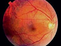
Diabetic Retinopathy.

Diabetic retinopathy is the leading cause of new blindness in persons aged 25-74 years in the United States. Approximately 700,000 persons in the United States have proliferative diabetic retinopathy, with an annual incidence of 65,000. A recent estimate of the prevalence of diabetic retinopathy in the United States showed a high prevalence of 28.5% among those with diabetes aged 40 years and older


http://img.medscape.com/pi/emed/ckb/endocrinology/1189694-1225122-41tn.jpg
Patients with diabetes often develop ophthalmic complications, such as corneal abnormalities, glaucoma, iris neovascularization, cataracts, and neuropathies. The most common and potentially most blinding of these complications, however, is diabetic retinopathy
In the initial stages of diabetic retinopathy, patients are generally asymptomatic, but in more advanced stages of the disease patients may experience symptoms that include floaters, distortion, and/or and blurred vision. Microaneurysms are the earliest clinical sign of diabetic retinopathy


 The exact mechanism by which diabetes causes retinopathy remains unclear
Of the approximately 16 million Americans with diabetes, 50% are unaware that they have it. Of those who know they have diabetes, only half receive appropriate eye care. Thus, it is not surprising that diabetic retinopathy is the leading cause of new blindness in persons aged 25-74 years in the United States.

Understanding Proliferative Diabetic Retinopathy

Diabetic Retinopathy


Diabetes is responsible for approximately 8000 eyes becoming blinded each year, meaning that diabetes is responsible for 12% of blindness. The rate is even higher among certain ethnic groups. An increased risk of diabetic retinopathy appears to exist in patients of Native American, Hispanic, and African American heritage
==================

JOIN OUR PAGE IN FACEBOOK
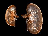
Diabetic Nephropathy

Diabetic nephropathy is a clinical syndrome characterized by the following:

Try Audible and Get Two Free Audiobooks
http://photos3.fotosearch.com/bthumb/ULY/ULY077/u11650419.jpg
Persistent albuminuria (>300 mg/d or >200 μg/min) that is confirmed on at least 2 occasions 3-6 months apart
Progressive decline in the glomerular filtration rate (GFR)
Elevated arterial blood pressure

Eat to Live: The Amazing Nutrient-Rich Program for Fast and Sustained Weight Loss


Currently, diabetic nephropathy is the leading cause of chronic kidney disease in the United States and other Western societies. It is also one of the most significant long-term complications in terms of morbidity and mortality for individual patients with diabetes. Diabetes is responsible for 30-40% of all end-stage renal disease (ESRD) cases in the United States.


Generally, diabetic nephropathy is considered after a routine urinalysis and screening for microalbuminuria in the setting of diabetes. Patients may have physical findings associated with long-standing diabetes mellitus.
Good evidence suggests that early treatment delays or prevents the onset of diabetic nephropathy or diabetic kidney disease
================
JOIN OUR PAGE IN FACEBOOK
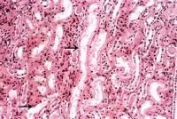
Acute Renal Faliure (Kidney Failure)

Acute renal failure (ARF), or acute kidney injury (AKI), as it is now referred to in the literature, is defined as an abrupt or rapid decline in renal filtration function. This condition is usually marked by a rise in serum creatinine concentration or by azotemia (a rise in blood urea nitrogen [BUN] concentration)
-->

http://img.medscape.com/pi/emed/ckb/nephrology/238062-243492-3329tn.jpg
However, immediately after a kidney injury, BUN or creatinine levels may be normal, and the only sign of a kidney injury may be decreased urine production

A rise in the creatinine level can result from medications (eg, cimetidine, trimethoprim) that inhibit the kidney’s tubular secretion. A rise in the BUN level can occur without renal injury, resulting instead from such sources as GI or mucosal bleeding, steroid use, or protein loading, so a careful inventory must be taken before determining if a kidney injury is present

Acute Renal Failure Part 1

Peritoneal Dialysis in Renal faluire
 
Hemodialysis Animation
 

==================== 


JOIN OUR PAGE IN FACEBOOK
======================

Join Our Channel in Youtube
Medical Learninig Videos

 Acute Renal Failure Part 2

-->

Status Epilepticus: Causes and Management

Status epilepticus (SE) is a common, life-threatening neurologic disorder. It is essentially an acute, prolonged epileptic crisis
causes:


Stroke (remote or acute)
Hypoxic injury
Tumor
Subarachnoid hemorrhage
Head trauma
Drugs (eg, cocaine, theophylline); isoniazid (INH) may cause seizures and is unique in having a specific antidote, pyridoxine (vitamin B-6)
Alcohol withdrawal
Electrolyte abnormalities (eg, hyponatremia, hypernatremia, hypercalcemia, hepatic encephalopathy)
Neoplasms
CNS infections (eg, meningitis, brain abscess, encephalitis)
Toxins, notably sympathomimetics

Antiepileptic Drug:
Benzodiazepines are the preferred first-line agents. Although diazepam is familiar to paramedics and emergency physicians, a consensus has evolved among neurologists and epileptologists that lorazepam may be preferred in this setting because of its long distribution half-life.


A comparison of initial IV treatment for overt generalized convulsive SE by Treiman et al found that lorazepam was more effective than phenytoin alone. Lorazepam was not more effective than phenobarbital or diazepam plus phenytoin, but it was easier to use. Not studied was fosphenytoin, which is theoretically a significant improvement over phenytoin.


Intravenous valproic acid has been shown in a pilot study to be equal to or better than phenytoin in aborting generalized SE, and it has been used in some cases of focal status epilepticus.


The use of levetiracetam (Keppra) in treatment of refractory SE has been examined, in part due to its availability in intravenous form, although its use in treating focal SE remains investigational. Anecdotal reports describe the beneficial use of topiramate in some cases of focal SE.

First Aid for Status Epilepticus

There are several intravenous formulations of antiepileptic drugs (AEDs) at different stages of development. Some of these might be able to help refractory cases with SE as adjunctive therapy.


No data clearly support a best third-line drug. Controlled trials are lacking, and recommendations vary greatly. While phenobarbital has historically been among the most widely used, the list of third-line drugs also includes midazolam, propofol, pentobarbital, valproate, levetiracetam, lidocaine, and others. Lacosamide, a novel antiepileptic drug available for intravenous injection, may be used safely as adjunctive therapy for SE, but little data exist on its efficacy.


A clinical practice trend seems to be for use of propofol as a third-line agent, often initiated during induction for endotracheal intubation. However, propofol infusion syndrome and increased mortality is reported when used at high doses and for prolonged periods. 
=========================

JOIN OUR PAGE IN FACEBOOK

 
Design by Free WordPress Themes | Bloggerized by Lasantha - Premium Blogger Themes | cheap international calls