Saturday, August 27, 2011

Differences Between Subdural and Epidural Hematomas


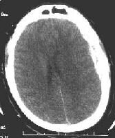
  first define the term "subdural hematoma " as it is the most common of the two types of hematoma . Subdural hematomas are essentially a pooling or collection of blood between the dura (the brain's protective covering, or outer layer) and a part of the meningies known as the arachnoid. What this means is that blood is pooling and then settling on the brain's fragile tissue. Depending on the severity of the bleed and the pressure, this can be extremely serious and even life threatening. Typically, subdural hematomas are caused by head trauma of some kind like a car accident or violent movement like a shaken baby would experience. What most often happens is that a small group of veins near the brain, called bridging veins, tear or shear due to head trauma or violent shaking. Since these veins tear, they allow blood to seep out into the space between the dura and the meningies, causing what is called intercranial pressure (pressure on the brain).


Other causes of subdural hematomas are use of medications that thin the blood called anticoagulants; long term aspirin and ibuprofen use cause the blood to be thin and therefore make bleeds more likely. The elderly are often candidates for subdural hematomas due to weaker blood vessels and sometimes brain atrophy. Brain atrophy means that the brain becomes smaller which causes a pulling on the bridging veins, followed by tears (almost like a run in a pair of lady's stockings).


The symptoms can include seizures, ataxia, lethargy, vomiting, visual problems, numbness, and ultimately coma and death. The best treatment for subdural hematomas are corticoid steroids to decrease swelling and surgery to alleviate pressure on the brain by draining some of the extra blood causing the pressure by drilling small holes in the cranium to release the access fluid.
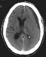
In contrast, an "epidural hematoma" is caused by arterial tears as opposed to the tearing or shearing of veins. Also, the epidural hematoma is located outside of the dura, between the dura and the cranium. Epidural hematomas are often caused by head trauma, more specifically a blow to the side of the head that causes a fracture in the skull or temporal bone. Less than a third of epidural hematomas occur outside of the area of where the temporal bone is located. Epidural hematomas can be very dangerous and also life threatening, as about one in every five cases of people treated for the condition dies. Because epidural hematomas are essentially arterial bleeds, there is a greater volume of blood loss in a shorter period of time (arteries are larger than veins with larger blood volumes). As the intercranial cavity fills with blood, the dura can often strip away from the brain causing an extremely painful headache. This bleed from arteries can bring on symptoms much more quickly as the pressure increases more rapidly than with a subdural hematoma.

Epidural hematomas can also occur in the areas near the spinal cord as a rare complication from anesthesia or other surgeries. The treatment for epidural hematomas is a quick diagnosis (often during a lucid period experienced by the patient, a conscious state between states of unconsciousness which is a key characteristic of hematomas) followed by neurosurgery. Again, it is necessary to drill small holes into the cranium in order to relieve pressure on the fragile tissue of the brain. Without this surgery, death is eminent.


we hope that our sit usefull for you?

Sited:
www.nlm.nih.gov/medlineplus/enc/article000781.htm
en.wikipedia.org/wik/Epidural_hematoma
===============

JOIN OUR PAGE IN FACEBOOK

Subdural Haematoma Surgery Video


A subdural hematoma (SDH) is a common neurosurgical disorder that often requires surgical intervention. It is a type of intracranial hemorrhage that occurs beneath the dura (essentially, a collection of blood over the surface of the brain) and may be associated with other brain injuries.
A left-sided acute subdural hematoma (SDH). Note tA left-sided acute subdural hematoma (SDH). Note the high signal intensity of acute blood and the (mild) midline shift of the ventricles. A left-sided chronic subdural hematoma (SDH). NoteA left-sided chronic subdural hematoma (SDH). Note the effacement of the left lateral ventricle. 

subdural hematoma evac burr holes


====================

JOIN OUR PAGE IN FACEBOOK

CT Scan of the Head video


CAT is very important in this section in comparison with other parts in the body,so we can not imagine any neuro-surgeon not use CAT.
This video for CAT Brain and Skull
This video for Normal CT


Subarachnoid hemorrhage

Acute subdural haematoma


Epidural Hematoma


Epidural Hematoma & Subdural Hematoma


CT Scan Abdominal Colonoscopy with Diabetic patient video


  CT for abdomen less important in comparison with CT for brain or skull especially with head trouma during RTA or Fall down from height

Instructions for Diabetics

  • Insulin:  If you are receiving insulin as part of your diabetes regimen, every attempt will be made to schedule your CT scan between 7:30 and 9:00 am.  Please consult with your physician regarding the dose of insulin you need to take before the CT scan.  Inform your physician that you cannot eat any solid food for 4 hours prior to the scan.
  • Metformin (Glucophage®): Do not take metformin on the day of your CT scan and do not resume taking it until your doctor has instructed you to do so.  Note: Metformin is also present within drugs named Glucovance, Avandamet, Metaglip, Fortamet, and Riomet.  After the CT scan, you must have another blood test to check your kidney function before you can start taking metformin again. Call your doctor for the results of the blood test for kidney function and for instructions about resuming metformin.  This is to prevent kidney damage and a serious reaction called lactic acidosis. 


On the Day of the CT Scan 

  • Do not eat any solid food for 4 hours prior to your CT scan.  You may drink clear liquids up until the time of your scan.  Clear liquids include water, black coffee or tea, apple juice, clear soda or clear broth.
  • Take your regular medications at your usual time, except insulin or metformin (see Instructions for Diabetics on other side).
  • Start drinking the oral contrast 1 ½ to 2 hours before your CT scan.  It can take from 30 to 60 minutes to drink all the contrast; you will need to be finished drinking all the oral contrast 1 hour before your scan.

During the CT Scan

You will lie on a table that will pass slowly through a large opening in the scanner as x-rays are taken.  You will be asked to lie perfectly still throughout the procedure, so that blurring does not occur.  Even though you will be alone in the room, you will be closely observed at all times.  If contrast is used, it will be injected into your arm through an IV line.  At the time of injection, you may have a momentary feeling of warmth and flushing, a salty taste in your mouth, and possibly some mild nausea.  This should pass quickly.  The scan itself is painless and should take approximately 30 minutes.
 video ct abdomen without contrast

After the CT Scan

After the scan, you should be able to resume your normal diet and activities.  Drink at least 5 to 6 glasses of water a day for 2 days after the scan. The water helps flush the contrast media from your system.  If you must limit your fluid intake because of a heart problem or for any other reason, talk with your doctor about how much water you can safely drink.
If you are a diabetic who takes any medication that contains metformin, you must have a blood test to check your kidney function before you can start taking metformin again. Call your doctor for the results of the blood test and for instructions about resuming metformin.  This is to prevent kidney damage and a serious reaction called lactic acidosis.
source: http://health.ucsd.edu/specialties/radiology/ct/abdomen.htm

Abdominal and Pelvic CT


A 3D reconstructed CT view of the kidneys and ureters, which connect the kidneys to the bladder. Part of the ribs, spine and pelvis are included in this image. 
CT scanning: sometimes called CAT scanning is a noninvasive medical test that helps physicians  to diagnose and treat medical diseases
CT scanning combines special x-ray equipment with sophisticated computers to produce multiple images or pictures of the inside of the body. These cross-sectional images of the area being studied can then be examined on a computer monitor, printed or transferred to a CD.
CT scans of internal organs, bones, soft tissue and blood vessels provide greater clarity and reveal more details than regular x-ray exams.
 
The video will describe anatomical structures as seen on a CT Scan

3D abdominal view 

3D anatomical view of the liver, vessels, tumor, kidneys, bones, extracted from CT scan images

Abdominal 3D viewing

A three-dimensional rendering of a mult-slice Computed Tomography dataset.
Rendering on a 3vision workstation


Computed Tomography CT video


CT Is important methods of radiology in making diadnosis of many disease so we must take some data about the CT.follow us in this department and we promise that you will be good radiologist
In a conventional x-ray structures are superimposed on top of one another, making it difficult to get a clear picture. Computed tomography, or CT scans take a picture of a single cross-section of the body, giving radiologists an unobstructed view of every organ, bone and soft-tissue. CT uses pencil-point x-ray beams from a rotating doughnut surrounding the patient's body. Detectors pick up the signals which vary in strength after they pass through human tissue. The detectors then send this information to a computer, which creates a picture of a single slice of the human body. A picture that looks something like—a pizza.

follow us in this blogger

Friday, August 26, 2011

How the MRI works


The majority of Medical student and some Doctors conflict about MRI so we want to give some data about MRI and videos to give some knowlege
http://www.radiologyinfo.org/photocat/thumbnail/philips6.jpg
Unlike conventional x-ray examinations and computed tomography (CT) scans, MRI does not depend on ionizing radiation. Instead, while in the magnet, radio waves redirect the axes of spinning protons, which are the nuclei of hydrogen atoms, in a strong magnetic field.
The magnetic field is produced by passing an electric current through wire coils in most MRI units. Other coils, located in the machine and in some cases, placed around the part of the body being imaged, send and receive radio waves, producing signals that are detected by the coils.
A computer then processes the signals and generates a series of images each of which shows a thin slice of the body. The images can then be studied from different angles by the interpreting physician.
Overall, the differentiation of abnormal (diseased) tissue from normal tissues is often better with MRI than with other imaging modalities such as x-ray, CT and ultrasound.

MRI of Brain


3D MPRAGE MRI scan acquired with a 32 channel head coil on a 3 Tesla Siemens Trio scanner at MGH in Boston

Indication

MR imaging of the Brain is performed to help diagnose:
  • tumors of the brain Benign or malignant
  • developmental anomalies of the brain appeared clinically or by CT
  • vascular anomalies of the head (aneurysm for example).
  • stroke.
  • trauma patients (in selected patients).
  • disease in the pituitary gland.
  • certain chronic disorders of the nervous system, such as multiple sclerosis.
  • disorders of the eyes and the inner ear.
  • causes of persistant headache.
Physicians also use the MR examination to document brain abnormalities in patients with dementia.
see this video


Anatomt of Brain Video


Here  we will give you a small hint about Brain anatomy by video so we will give you introduction
http://photos2.fotosearch.com/bthumb/VSL/Anatomy/094_1866.jpg


befor video
The anatomy of the brain is complex due its complex structure and function. This  organ acts as a control center by receiving, interpreting, and directing sensory information throughout the body. There are three major divisions of the brain. They are the forebrain, the midbrain, and the hindbrain
 Now you can see the video


3D MRI Human Brain Anatomy

This is a technique we developed called deep rendering in an effort to create an application that would allow users to explore the interior of the human body, in real-time. For more information

BUY NOW and save 21$
Acland's DVD Atlas of Human Anatomy, Set of Six DVDs: The Upper Extremity, The Lower Extremity, The Trunk, The Head and Neck, Part 1, The Head and Neck, Part 2, and The Internal Organs  


Design by Free WordPress Themes | Bloggerized by Lasantha - Premium Blogger Themes | cheap international calls